
Quick Take
- CLL diagnosis starts with a complete blood count and peripheral blood smear.
- Flow cytometry confirms the immunophenotype of malignant B‑cells.
- FISH, TP53 mutation analysis, and IGHV status guide prognosis.
- Bone‑marrow biopsy is reserved for atypical cases or when staging requires it.
- Rai and Binet staging translate test results into treatment decisions.
What Is Chronic Lymphocytic Leukemia?
Chronic lymphocytic leukemia is a slow‑growing malignancy of mature B‑lymphocytes that primarily accumulates in the blood, bone marrow, and lymphoid tissues. It accounts for about 25% of adult leukemias in Western countries, with a median diagnosis age of 70 years. The disease’s hallmark is an absolute lymphocytosis-usually >5×10⁹/L-combined with characteristic immunophenotypic markers (CD5, CD19, CD23) on flow cytometry.
Core Blood Tests: CBC and Peripheral Blood Smear
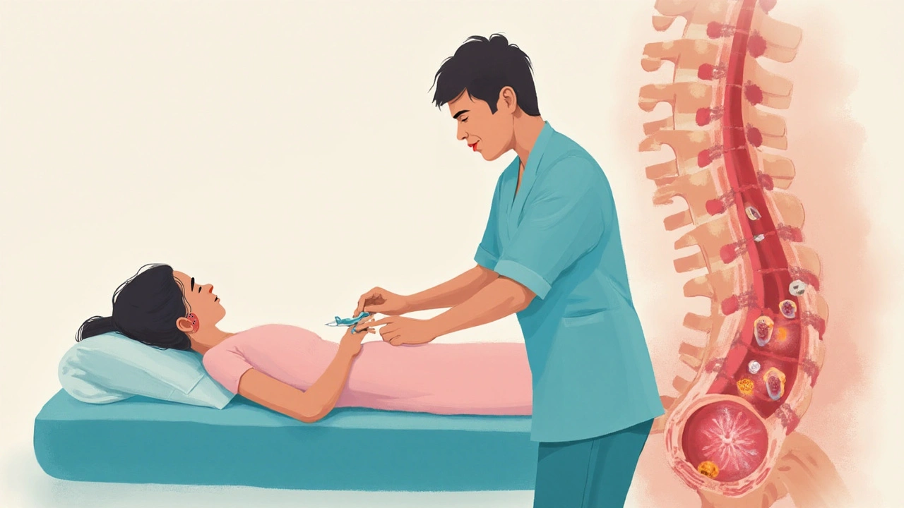
The first clue appears in a complete blood count (CBC). A CBC provides quantitative data: hemoglobin, platelets, and a white‑blood‑cell differential. In CLL, the total leukocyte count is often elevated, driven by a high lymphocyte percentage.
Next, a peripheral blood smear is examined under a microscope. The smear reveals small‑to‑medium‑sized lymphocytes with a "smudge cell" appearance-fragile cells that rupture during slide preparation. These morphological clues raise suspicion but are not definitive.
Flow Cytometry and Immunophenotyping
The gold‑standard confirmation comes from flow cytometry. This technique tags cells with fluorescent antibodies targeting surface markers. In CLL, the pattern typically shows CD5⁺, CD19⁺, CD20⁺ (dim), CD23⁺, and weak surface immunoglobulin. The test’s sensitivity exceeds 95%, making it indispensable for distinguishing CLL from other lymphoproliferative disorders.
Immunophenotyping results are recorded as a profile of antigen expression. When the profile deviates-e.g., strong CD20 or CD200 absence-physicians may explore mantle‑cell lymphoma or monoclonal B‑cell lymphocytosis as alternatives.
Cytogenetic and Molecular Tests
Genetic abnormalities heavily influence prognosis. The most common assay is fluorescence in‑situ hybridisation (FISH), which detects deletions at 13q14, trisomy 12, 11q22‑23 (ATM), and 17p13 (TP53). A 17p deletion or TP53 mutation signals high‑risk disease, often prompting early treatment.
Beyond FISH, targeted sequencing evaluates TP53 mutation. Even in the absence of a 17p deletion, a TP53 point mutation carries a similarly poor outlook.
Another molecular hallmark is the IGHV mutational status. Unmutated IGHV (<98% germline identity) correlates with aggressive disease, while mutated IGHV suggests a more indolent course.

Bone Marrow Evaluation
While many CLL cases are diagnosed from blood alone, a bone‑marrow biopsy may be necessary when lymphocytosis is borderline, when cytopenias develop without clear cause, or when histologic confirmation is required for clinical trials. The biopsy provides cellularity data, infiltration patterns, and a substrate for additional cytogenetic studies.
Biopsy specimens are also examined with flow cytometry, ensuring concordance between peripheral and marrow compartments.
Staging Systems: Rai and Binet
After diagnostic confirmation, clinicians assign a stage to predict survival and guide therapy. The Rai staging system (used primarily in the United States) categorises patients into low (stage 0), intermediate (stage I-II), and high (stage III-IV) based on lymphadenopathy, organomegaly, anemia, and thrombocytopenia.
In Europe, the Binet classification splits patients into A, B, or C groups depending on the number of involved lymphoid sites and the presence of cytopenias. Both systems incorporate laboratory findings from the CBC, marrow, and cytogenetic tests.
Imaging and Ancillary Tests
When physical examination reveals bulky lymphadenopathy or organomegaly, imaging such as a contrast‑enhanced computed tomography (CT) scan clarifies disease extent. Positron emission tomography (PET) is less routine but may be used to differentiate Richter transformation-an aggressive evolution of CLL into diffuse large B‑cell lymphoma.
Serum immunoglobulin levels (IgG, IgA, IgM) are also measured; hypogammaglobulinemia occurs in up to 50% of patients and predisposes to infections, influencing prophylactic strategies.
Putting It All Together: Diagnostic Algorithm
Below is a concise flow of how the tests interlock:
| Test | Primary Insight | Sensitivity | Invasiveness |
|---|---|---|---|
| CBC & Peripheral Smear | Quantifies lymphocytosis; identifies smudge cells | ≈70‑80% | Non‑invasive |
| Flow Cytometry | Confirms CLL immunophenotype | >95% | Minimally invasive (blood draw) |
| FISH Cytogenetics | Detects high‑risk chromosomal lesions | ≈90% | Minimally invasive (blood or marrow) |
| Bone Marrow Biopsy | Assesses marrow infiltration, provides tissue for cytogenetics | ≈85% | Invasive (needle core) |
In practice, a CBC and smear trigger flow cytometry. If flow confirms CLL, FISH (plus TP53 sequencing) follows. Bone‑marrow biopsy is added only when indicated by atypical blood results or when a clinical trial requires it.
Related Concepts
Understanding CLL diagnosis opens doors to adjacent topics such as targeted therapies (BTK inhibitors, BCL‑2 antagonists), prophylactic immunoglobulin replacement, and the management of Richter transformation. Readers interested in treatment pathways can explore the "Therapeutic Options for CLL" article, while those focusing on patient monitoring may wish to read about "Long‑Term Surveillance Strategies".
Frequently Asked Questions
Can CLL be diagnosed without a bone‑marrow biopsy?
Yes. In most cases, a peripheral blood smear combined with flow cytometry and FISH provides enough information for a definitive diagnosis. Biopsy is reserved for atypical presentations or research protocols.
What does a 17p deletion mean for a CLL patient?
A deletion of chromosome 17p includes the TP53 gene, which is a key tumor‑suppressor. Its loss predicts resistance to standard chemoimmunotherapy and often leads clinicians to choose targeted agents like ibrutinib or venetoclax earlier.
How often should a patient with early‑stage CLL undergo repeat testing?
Guidelines recommend CBC and clinical review every 3-6months for low‑risk disease. Flow cytometry and FISH are typically repeated only if there’s a clinical change, such as new lymphadenopathy, cytopenias, or a rapid rise in lymphocyte count.
Is IGHV mutational status used to decide when to start treatment?
IGHV status alone does not trigger therapy, but an unmutated IGHV score (≤98% similarity to germline) signals a higher likelihood of disease progression. Physicians combine this information with staging, symptoms, and cytogenetics to time treatment.
Do smudge cells on a blood smear definitively indicate CLL?
No. Smudge cells are characteristic but not exclusive to CLL; they can appear in other lymphoproliferative disorders and even in healthy older adults. Confirmation requires flow cytometry.
Stephanie Zuidervliet
September 24, 2025 AT 10:07Wow!!! This article just blew my mind... the cascade of tests, the flow cytometry drama, the FISH fireworks!!! It's like a medical thriller, but with more punctuation!!! But seriously, who has time for all these labs?
Olivia Crowe
September 24, 2025 AT 13:12Great overview-keep shining!
Aayush Shastri
September 24, 2025 AT 16:17As someone from the subcontinent, I appreciate the clear breakdown, especially the emphasis on cost‑effective peripheral smear before jumping to invasive biopsies. It respects resource‑limited settings while staying scientifically rigorous.
Quinn S.
September 24, 2025 AT 19:22While the article is generally accurate, it suffers from a lack of precision in describing the sensitivity of flow cytometry; the claim of >95% should be qualified with methodological caveats, and the discussion of TP53 mutation analysis is overly simplistic.
Dilip Parmanand
September 24, 2025 AT 22:27Spot on-lab work can feel endless.
Sarah Seddon
September 25, 2025 AT 01:33Reading this felt like watching a vibrant kaleidoscope of diagnostic tools! The way you painted the picture of smudge cells dancing on the slide was nothing short of poetic, and the guidance on when to biopsy is a lifesaver for clinicians.
Ari Kusumo Wibowo
September 25, 2025 AT 04:38Yo, love the vibe, but cut the fluff-just tell us when to order FISH and we’re good.
Hannah Gorman
September 25, 2025 AT 07:41First, let me commend the thoroughness of the piece; it covers the entire diagnostic algorithm without omitting any critical step. The author starts with the obvious CBC, which is a staple in any hematologic workup, and correctly notes the significance of lymphocytosis. Moving on, the description of smudge cells is accurate, though it could benefit from a brief mention of their occasional presence in healthy elderly individuals. Flow cytometry is rightly highlighted as the definitive test, and the antigen panel is spot on. However, the piece glosses over the nuances of CD20 intensity, which can sometimes mislead diagnosis. The discussion on cytogenetics, particularly FISH for 13q14 deletion, is essential for risk stratification. I appreciate the emphasis on TP53 mutation analysis, a predictor of poor response to chemoimmunotherapy. The IGHV mutational status section is well placed, but a deeper dive into its impact on disease kinetics would enrich the narrative. The author wisely cautions that bone‑marrow biopsy is not routine, reserving it for atypical presentations. This aligns with current guidelines that prioritize minimally invasive approaches. The staging systems, Rai and Binet, are mentioned, yet the interplay between staging and molecular findings could be more explicit. The FAQs are helpful, especially the clarification that smudge cells are not pathognomonic. Overall, the article balances brevity with depth, making it suitable for both trainees and seasoned clinicians. Still, a few minor edits-such as consistent use of abbreviations and clearer formatting of tables-would polish the final product.
Tatiana Akimova
September 25, 2025 AT 10:30Don't let all these acronyms intimidate you-master them one step at a time, and you'll become the diagnostic hero your patients need!