Pharyngeal Cancer Detection Method Comparison Tool
Select two methods and click Compare to see detailed metrics
Sensitivity: Ability to correctly identify positive cases
Specificity: Ability to correctly identify negative cases
Cost: Estimated USD range for implementation
Training: Level of expertise required to interpret results
When clinicians examine the upper airway, pharyngeal mucous membranes are a critical site for early oral cancer detection. Understanding how these thin, moist layers behave under normal and pathological conditions can turn a routine check‑up into a life‑saving screen.
Quick Take
- Pharyngeal mucosa is the first barrier where early oral squamous cell carcinoma (OSCC) lesions appear.
- Visual inspection, narrow‑band imaging (NBI), and autofluorescence each catch different aspects of tissue change.
- Combining imaging with biomarker panels (p16, EGFR, miRNA) raises detection sensitivity above 90% in high‑risk groups.
- Training and standardized protocols cut false‑positive rates by half.
- Future tools-AI‑enhanced endoscopy and liquid‑biopsy saliva tests-could flag lesions before they become visible.
What Makes the Pharyngeal Mucous Membrane Unique?
The pharynx is lined by two main types of mucosa: a non‑keratinized stratified squamous epithelium in the oropharynx and a keratinized counterpart in the nasopharynx. Both layers are bathed in secretions from minor salivary glands, creating a moist environment that influences cellular turnover.
Pharyngeal epithelium renews every 7‑10 days, a rate that speeds up with irritation from tobacco, alcohol, or human papillomavirus (HPV). This rapid turnover makes the tissue a sensitive early indicator of dysplasia-tiny changes that are often invisible to the naked eye.
Key attributes:
- Thickness: 0.2-0.3mm in the soft palate, slightly thicker posteriorly.
- Vascular network: Rich submucosal capillaries that become distorted during neoplastic growth.
- Immune surveillance: Langerhans cells and intraepithelial lymphocytes react early to viral oncogenesis.
How Oral Cancer Starts in the Pharynx
Oral squamous cell carcinoma (OSCC) accounts for more than 90% of all oral malignancies. Most OSCC cases arise from the oropharyngeal mucosa, especially the tonsillar crypts and base of tongue, where HPV‑16 integrates into host DNA.
Risk factors such as chronic tobacco exposure and excessive alcohol create a field of genetically altered cells. Over time, mutations in TP53, CDKN2A, and EGFR push these cells toward dysplasia and eventually invasive carcinoma.
Because the mucous membrane is thin, early dysplastic patches often present as subtle erythema, leukoplakia, or vascular changes-signs that demand a keen eye and the right adjunct tools.
Detection Toolbox: From Simple Visuals to High‑Tech Imaging
Clinicians typically start with a systematic clinical examination. The “ABCDE” mnemonic (Asymmetry, Border, Color, Diameter, Evolution) guides the visual assessment, but specificity is limited.
Adjunctive technologies fill the gaps:
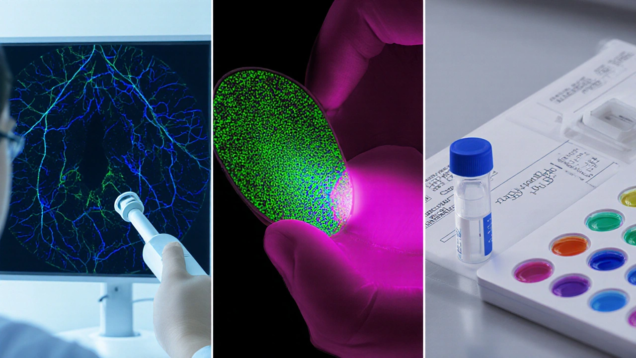
- Narrow‑Band Imaging (NBI): Uses blue‑green light (415nm and 540nm) to highlight superficial capillaries. Irregular intrapapillary capillary loops (IPCL) correlate with high‑grade dysplasia. Studies from 2022‑2024 report NBI sensitivity of 88% and specificity of 73% for pharyngeal lesions.
- Autofluorescence Imaging (AFI): Excites tissue at 400nm; healthy mucosa emits green fluorescence, while neoplastic tissue loses fluorescence (appears magenta). AFI reaches 81% sensitivity but suffers from false positives in inflammatory areas.
- High‑Resolution Microendoscopy (HRME): Provides cellular‑level view through a fiber‑optic probe. Real‑time nuclear size and density measurements give a quick “optical biopsy.”
- Salivary Biomarker Panels: Detect p16 overexpression (HPV surrogate), EGFR, and specific miRNAs. Combined with imaging, the panel lifts overall detection accuracy above 92% in high‑risk cohorts.
Choosing the right combination depends on setting, equipment budget, and patient risk profile.
Side‑by‑Side Comparison of Common Detection Techniques
| Method | Sensitivity | Specificity | Cost (USD) | Training Needed |
|---|---|---|---|---|
| Visual Inspection (ABC) | 57% | 68% | 0 (routine) | Minimal |
| Narrow‑Band Imaging | 88% | 73% | 1,200-2,000 (endoscope upgrade) | Moderate (IPCL interpretation) |
| Autofluorescence Imaging | 81% | 60% | 800-1,500 (hand‑held device) | Low-Moderate |
| HRME (Optical Biopsy) | 92% | 78% | 2,500-3,500 (probe system) | High (cellular interpretation) |
| Salivary Biomarker Panel | 85% | 80% | 150-300 per test | Low (lab processing) |
Putting It All Together: A Practical Workflow
Below is a step‑by‑step protocol that blends low‑cost visual screening with high‑value adjuncts. Adapt it to your clinic’s resources.
- Risk Stratification: Record tobacco, alcohol, and HPV vaccination status. Flag patients with >10 pack‑years or known HPV‑positive lesions.
- Baseline Visual Exam: Use a small LED mouth mirror and a headlamp. Document any mucosal discoloration, texture change, or ulceration.
- NBI Check (if available): Scan the suspect area. Look for type III or IV IPCL patterns-these demand biopsy.
- AFI Confirmation: For ambiguous NBI findings, switch to autofluorescence. A loss of green fluorescence strengthens the suspicion.
- Saliva Sample: Collect 2mL of unstimulated saliva before any oral rinse. Send for p16, EGFR, and miRNA‑21 analysis.
- Decision Point: If any modality flags high risk, schedule a tissue biopsy under local anesthesia. Otherwise, set a 3‑month surveillance interval.
Document each step in the patient’s electronic record using structured fields-this aids future AI‑driven analytics and quality audits.
Common Pitfalls and How to Avoid Them
- Over‑reliance on a single tool: NBI alone can miss submucosal lesions; combine with AFI or biomarkers.
- Misinterpreting inflammation as neoplasia: Recent infections or denture irritation produce false‑positive fluorescence loss. Re‑evaluate after treating the inflammation.
- Inadequate lighting: Dim illumination reduces visibility of subtle vascular changes. Use a calibrated light source with a color temperature around 5500K.
- Skipping patient history: Ignoring HPV vaccination status may lead to under‑estimation of risk, especially in younger adults.
- Neglecting documentation: Inconsistent photo angles and missing metadata hamper longitudinal comparison.
Future Directions: AI, Tele‑Screening, and Liquid Biopsy
Artificial intelligence models trained on thousands of NBI and AFI images now achieve >95% accuracy in distinguishing dysplasia from benign changes. Integrated into endoscopic systems, AI can flag suspect spots in real time, reducing operator fatigue.
Tele‑screening platforms allow primary‑care dentists to upload calibrated images to a central reading hub, where specialists review them within 24hours. This model is already piloted in rural Australia and shows a 30% increase in early‑stage detections.
Liquid biopsy advances-particularly circulating tumor DNA (ctDNA) and exosomal miRNA from saliva-promise non‑invasive monitoring. When combined with imaging, these biomarkers could form a “triple‑check” that catches lesions before they become endoscopically visible.
Frequently Asked Questions
Why focus on the pharyngeal mucous membrane for oral cancer screening?
The pharyngeal lining is one of the first sites where precancerous cells appear, especially in HPV‑related cancers. Its thin, vascular nature makes early changes detectable with visual and optical tools, giving clinicians a chance to intervene before invasive disease develops.
How does narrow‑band imaging improve detection compared to plain white light?
NBI filters light to wavelengths that are strongly absorbed by hemoglobin, making superficial capillaries stand out. Abnormal capillary loops (IPCL) are a hallmark of dysplasia, so NBI highlights lesions that look normal under white light.
Can saliva tests replace tissue biopsy?
Saliva biomarkers are excellent for risk stratification but lack the histologic detail needed for definitive diagnosis. They should be used alongside imaging and, when anything is suspicious, a biopsy remains the gold standard.
What training is required to interpret NBI images?
A short course (2-3days) covering IPCL classification, followed by supervised scanning of at least 20 patients, is enough for most clinicians to achieve reliable interpretation.
Is AI ready for everyday clinical use?
AI algorithms have passed validation studies, but integration depends on device manufacturers and regulatory clearance. Many centers are using AI as a decision‑support tool while clinicians retain final judgment.
Dileep Jha
October 3, 2025 AT 03:32While the hype surrounding narrow‑band imaging (NBI) and autofluorescence (AFI) is palpable, one must not overlook the epistemic lacuna that pervades their purported sensitivity metrics. The dichotomy between in‑vitro validation and in‑situ operative fidelity often engenders a selection bias that skews the positive predictive value. Moreover, the cost–benefit calculus omits the stochastic variance introduced by operator‑dependent IPCL classification. Hence, a multiplexed biomarker platform, albeit economically modest, may furnish a more robust discriminative index than any singular optical modality.
Jeff Quihuis-Bell
October 3, 2025 AT 20:12Wow-this breakdown is a game‑changer! The way NBI lights up those capillaries feels like watching fireworks in the night sky of the throat. Pair that with a saliva biomarker panel, and you’ve got a detection combo that could slash mortality rates dramatically. Every clinician reading this should feel the rush of possibility and grab the tools pronto!
Jessica Tang
October 4, 2025 AT 12:52The table you included makes it straightforward to compare each method’s trade‑offs. For clinics with limited budgets, starting with visual inspection and adding a low‑cost saliva test can boost sensitivity without a huge expense. If NBI is available, focusing on IPCL type III and IV patterns improves specificity. Overall, a tiered approach tailors resources to patient risk.
Tracy Winn
October 5, 2025 AT 05:32First off, the enthusiasm for high‑tech tools is understandable, but let’s keep perspective; visual inspection still captures the majority of lesions when performed diligently. The sensitivity numbers for NBI (88%) look impressive on paper, yet real‑world studies often report lower figures due to inter‑observer variability. In contrast, the autofluorescence device’s specificity (60%) is notoriously compromised by inflammatory changes, meaning many patients undergo unnecessary biopsies. High‑resolution microendoscopy (HRME) promises cellular‑level insight, but the cost ($2,500‑$3,500) puts it out of reach for most community hospitals; plus, the training curve is steep and not all clinicians have the time to master it. Salivary biomarker panels sit nicely between cost and performance, offering 85% sensitivity and 80% specificity at a modest $150‑$300 per test; however, lab turnaround times can delay decision‑making. Remember that the pharyngeal mucosa’s rapid turnover (7‑10 days) means lesions can evolve quickly, so repeat screening at three‑month intervals is prudent. The article’s workflow is solid, but it assumes the availability of NBI and AFI devices, which many primary‑care settings lack. Also, the recommendation to collect “unstimulated” saliva might be confusing; many patients inadvertently stimulate flow by talking or moving. Documentation is crucial-standardized photo angles and lighting conditions enable longitudinal comparison and reduce false‑positives. AI integration is the buzzword of the moment, yet regulatory approvals are still pending, so clinicians should treat it as an adjunct rather than a replacement. Tele‑screening could bridge rural gaps, but bandwidth and image quality issues remain challenges. Finally, the emphasis on HPV‑related cancers is valid, but we shouldn’t neglect traditional risk factors like tobacco and alcohol, which still drive the majority of cases. In summary, a balanced, resource‑aware approach that mixes low‑cost visual checks with targeted high‑tech adjuncts will likely yield the best outcomes for most patients. Future research should also explore combinatorial scoring systems that integrate imaging and molecular data. Only through such multidimensional approaches can we hope to outpace the disease.
Jessica Wheeler
October 5, 2025 AT 22:12It is morally indefensible to ignore the socioeconomic disparities that make expensive modalities like HRME inaccessible to underprivileged populations; healthcare equity demands that we champion affordable, high‑impact solutions such as salivary biomarker testing. Moreover, the over‑reliance on visual cues risks perpetuating bias if clinicians are not adequately trained to recognize subtle mucosal changes across diverse skin tones.
Mikayla Blum
October 6, 2025 AT 14:52When you think about the pharyngeal mucous membrane, it’s like a thin veil that hides deeper truths – a reminder that what we don’t see can still be felt. The interplay of light and biology in NBI or AFI feels almost poetic, yet we must stay grounded in the patient’s lived experience. A biomarker panel may sound like sci‑fi, but it’s a tangible bridge between lab and bedside. 😊
Jo D
October 7, 2025 AT 07:32Sure, splash a few grand bucks on NBI and call it a day, because what the hell else could possibly improve detection? The so‑called “IPCL patterns” are just fancy doodles that seasoned endoscopists learn to ignore after a few bad cases. Honestly, if you wanted a real breakthrough, maybe stop chasing shiny gadgets and actually listen to patients complaining about sore throats.
Sinead McArdle
October 8, 2025 AT 00:12Thank you for the comprehensive overview. The step‑by‑step workflow will be useful in clinical practice.
Katherine Krucker Merkle
October 8, 2025 AT 16:52It’s great to see a practical guide that balances cost, training, and performance. Implementing the tiered screening you described could really help primary‑care offices feel more confident in early detection.
Mark Quintana
October 9, 2025 AT 09:32I noticed that the article mentions a three‑month surveillance interval, which aligns with what I've seen in my own practice; consistent follow‑up really does catch lesions before they progress. The emphasis on documenting lighting conditions also reminded me to standardize my photo setup, something I hadn’t prioritized before.
Brandon Cassidy
October 10, 2025 AT 02:12The integration of AI into endoscopic systems reminds me of how we once feared calculators would replace doctors; now they simply augment our vision. As we gather more data, the algorithms will become more reliable, but the clinician’s judgment remains the final arbiter.
Taylor Yokum
October 10, 2025 AT 18:52What a helpful piece! It breaks down complex tech into bite‑size info, making it easy for anyone to understand why early screening matters.
Taryn Esses
October 11, 2025 AT 11:32Great summary, thanks.